Skip to content
Contact Us
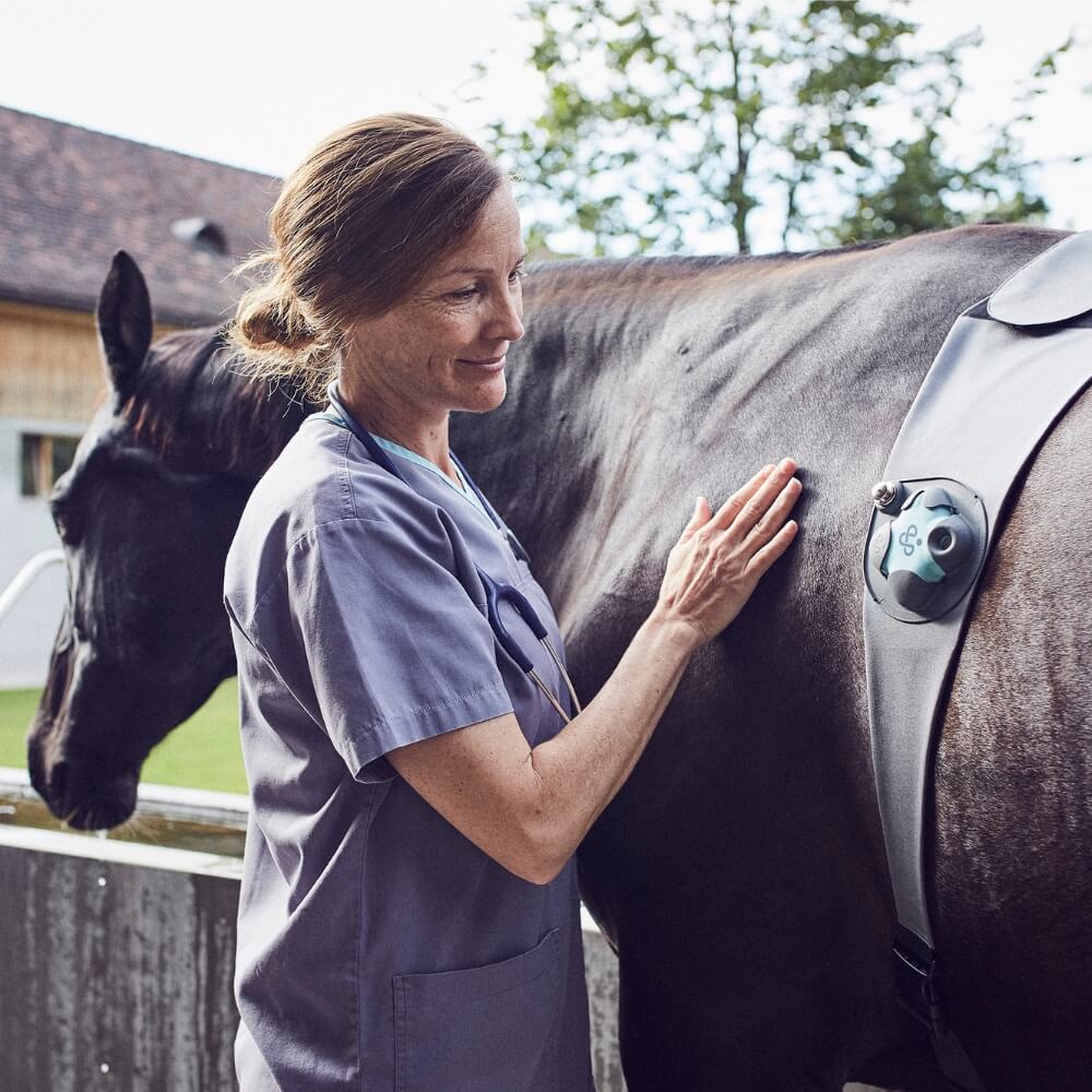
Setting the Standard in Digital Veterinary Care
Diagnostics
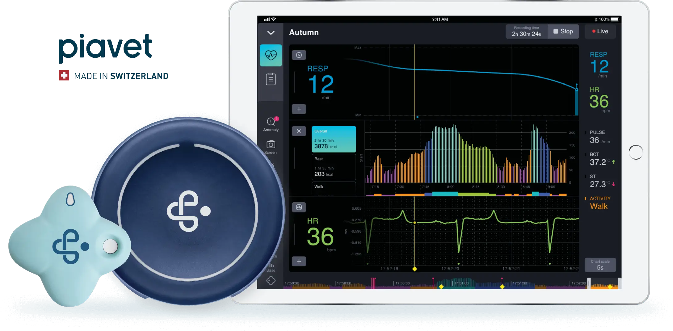
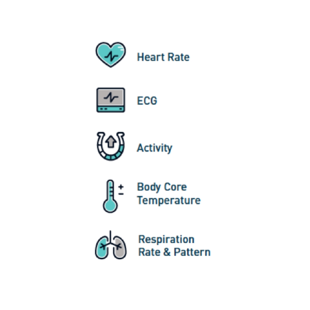
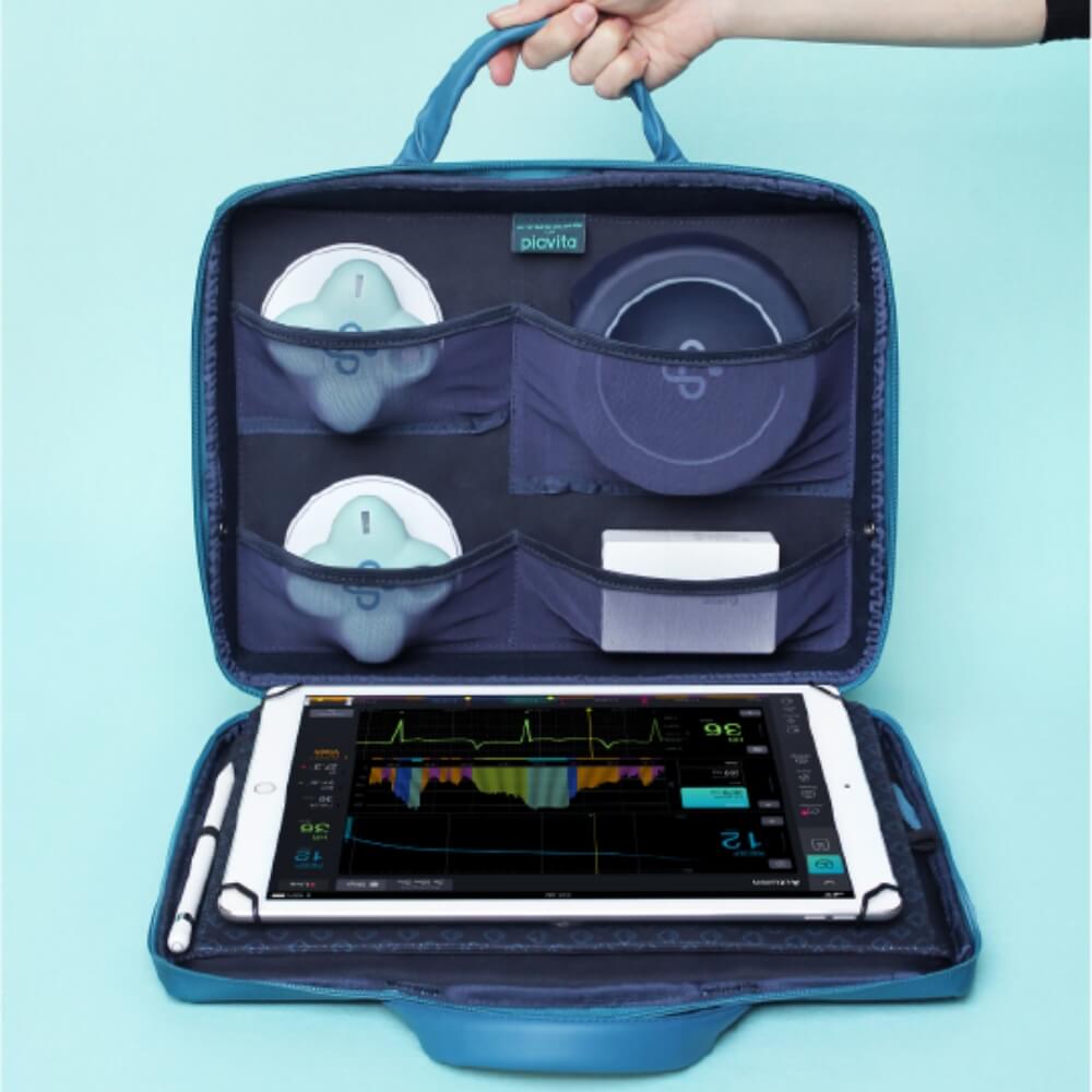
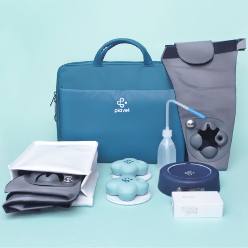
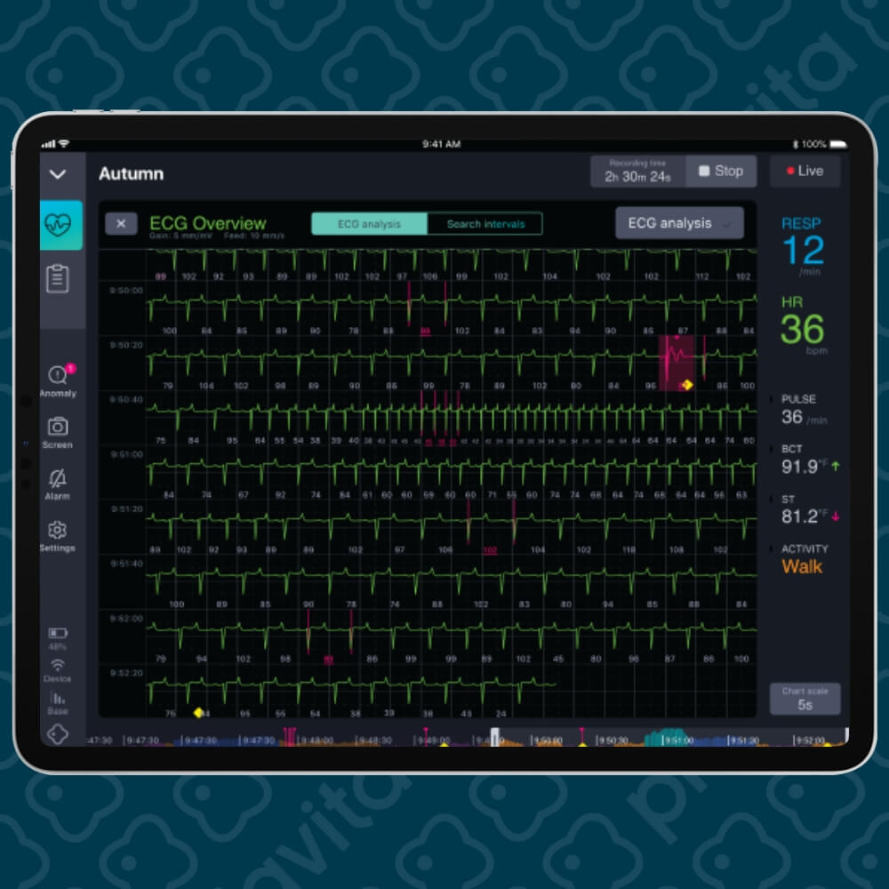
Vital Recording
Patient Monitoring
Patient Management
Predictive Health
Report Automation
AI – Smart Alerts
Schedule a Demo
Elevating Animal Health for Vets, Breeders, and Researchers: From driving Vets to pharmaceutical Researchers.
Vets & Breeders
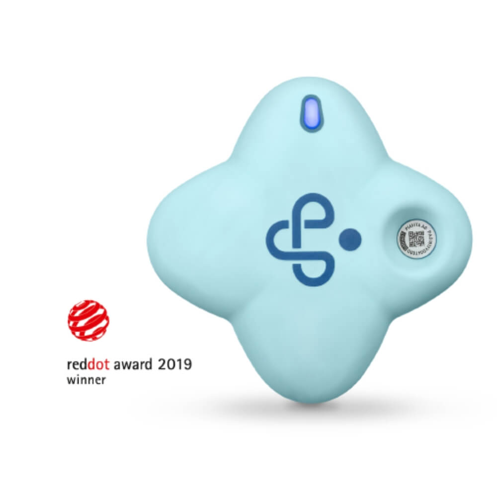
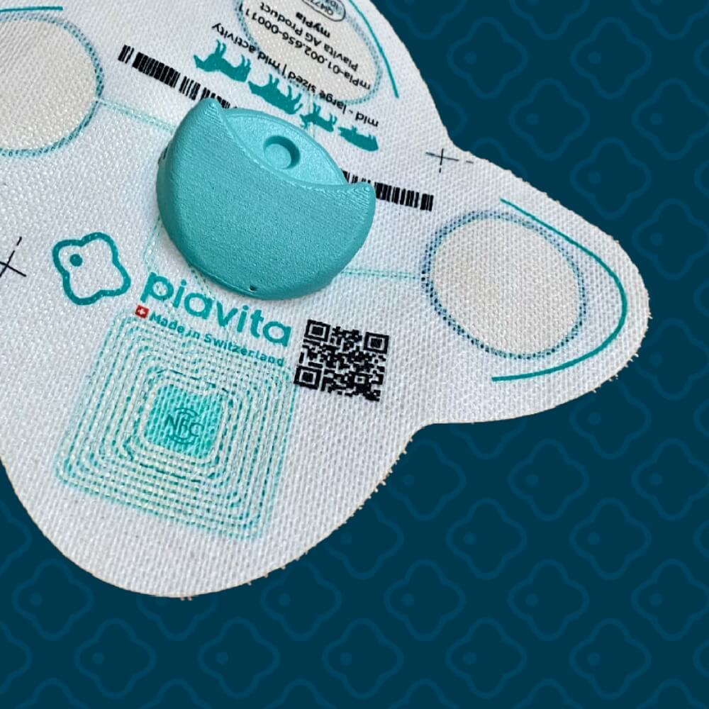
PiaVet
Schedule a Demo
Download Brochure
Researchers
PiaPatch
Get in Contact
Schedule a PiaVet Demo
Fill out the form below, and we will be in touch shortly.
Contact Information
Name
phone
email
Further information or questions
Message
I agree that my information in this contact form will be collected and processed to answer my request. I understand that I can revoke this consent at any time by sending an email to
[email protected]
. I found further information on how to handle user data in the
data protection declaration
.
submit ⟶
Contact Us
Fill out the form and we will be in touch shortly.
Name
Email
Message
I agree that my information in this contact form will be collected and processed to answer my request. I understand that I can revoke this consent at any time by sending an email to
[email protected]
. I found further information on how to handle user data in the
data protection declaration
.
submit ⟶